胎兒發育 產前檢查 盆腔超聲波 不育及輔助生殖 乳房檢查
Women's Health

產前檢查

Stages of Pregnancy 孕期

Pregnancy lasts about 40 weeks, counting from the first day of your last normal period. The weeks are grouped into three trimesters. Find out what checkups are to be done with you and your baby in these three stages.

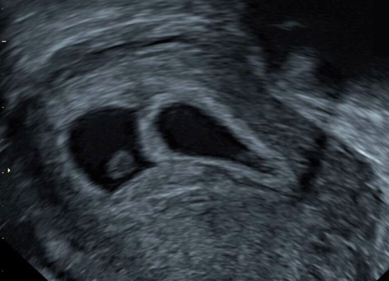
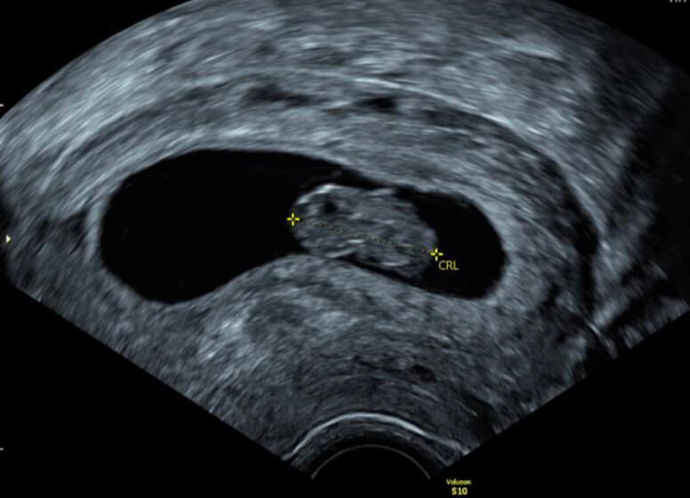
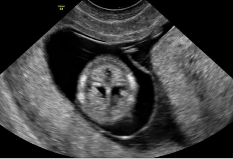
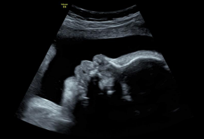
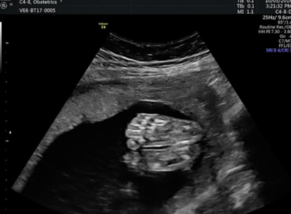
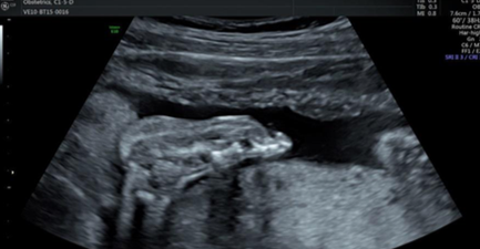
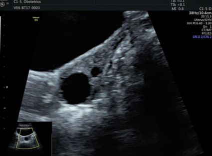
For many women, especially after 8 weeks gestation, sufficient information about the baby may be obtained with transabdominal ultrasound scan (經腹部超聲波掃描). Sometimes transvaginal ultrasound (經陰道超聲波掃描) may be required to get better and clearer images of the female pelvic organs including the developing pregnancy as the ultrasound probe lies closer to these organs.

WHAT TO BE CHECKED APART FROM ULTRASOUND:
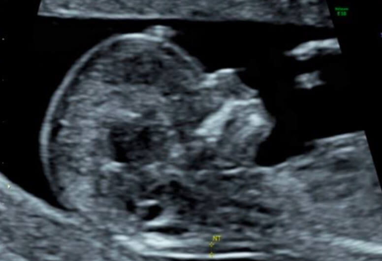
  • Sometimes the results of a first trimester ultrasound scan may be inconclusive or uncertain, and need to be combined with mother’s clinical history and other tests (e.g. blood tests such as NIPT, NIFTY, T21, Chorionic Villous Sampling 絨毛膜活檢, Amniocentesis 羊膜穿刺術, etc.) for an overall assessment.

NIPT (i.e. Non-Invasive Prenatal Test 無創性產前檢查)

can be done when gestational age is beyond 10 weeks. It works by testing mother’s blood, which also contain small amount of fetal DNA, for increased amount of DNA from Chromosome 21. Besides detecting Down syndrome, it can also detect Edward syndrome (an extra copy of Chromosome 18), Patau syndrome (an extra copy of Chromosome 13) and Turner syndrome (only 1 copy of Sex Chromosome [X]) and other chromosomal disorders. There are different types of NIPT, such as NIFTY and T21 test.

Chorionic Villous Sampling (絨毛膜活檢) , abbreviated “CVS”

involves getting a small sample of the placental tissue by a fine needle passing under ultrasound guidance through the mother’s tummy and her uterus to the placenta, where a small sample of tissue is obtained by suction. This procedure is an invasive one and carry a small miscarriage rate of 0.5-1% even conducted by experienced doctors. It is usually performed between 11-14 weeks of gestation.

Amniocentesis (羊膜穿刺術, 俗稱『抽羊水』)

involves withdrawing a small sample of amniotic fluid (羊水) from the mother’s uterus. Before the procedure, ultrasound scan is performed to locate the best site for sampling. During the procedure, a fine needle is passed through mother’s tummy into the amniotic cavity to withdraw a small amount of fluid. As amniotic fluid contains cells that the fetus has shed, it can be used for diagnosis. This is an invasive procedure carrying a small miscarriage rate of 0.5-1% even under experienced hands. It is usually performed between 16-20 weeks of gestation.

Morphology Ultrasound Scan

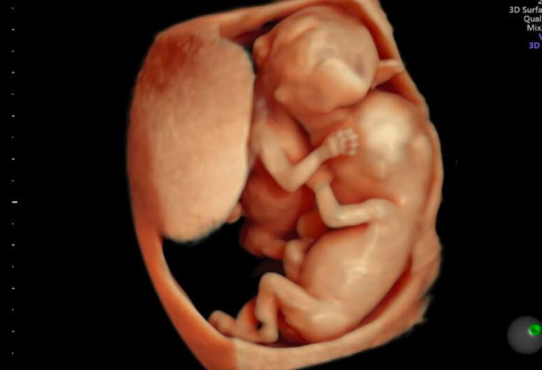
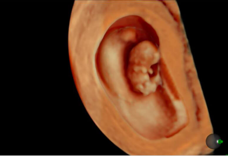
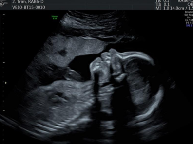
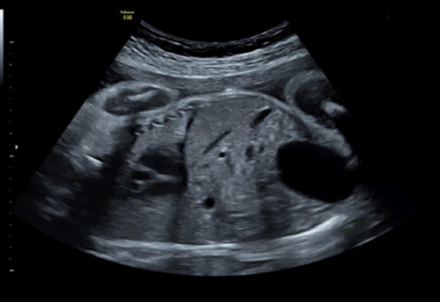
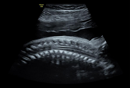
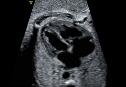
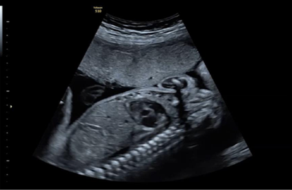
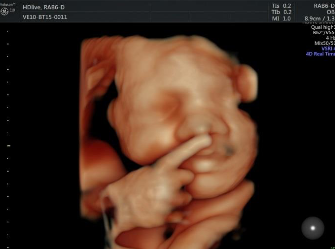
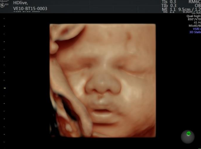
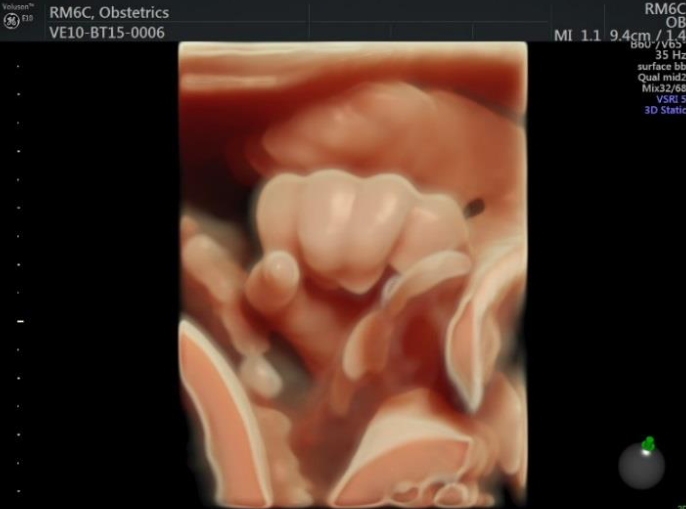
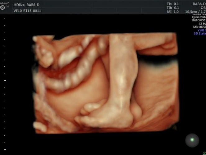
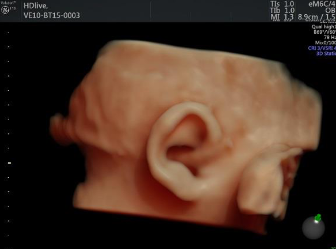
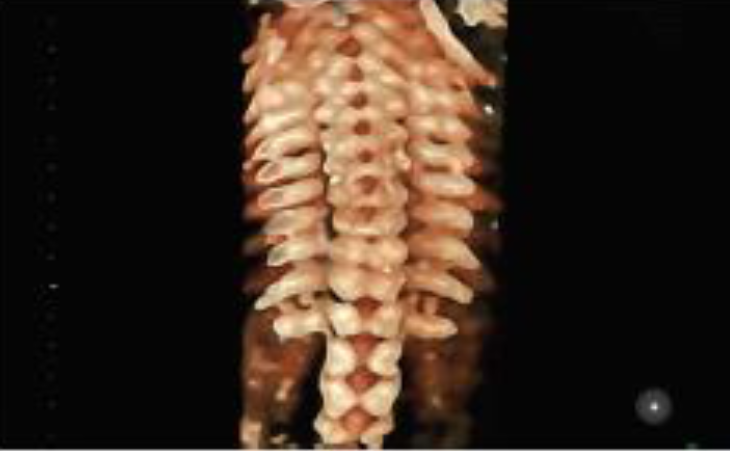
Fetal morphology ultrasound scan (胎兒結構性超聲波掃描), also called “structural scan” and “anomaly scan”, is usually performed during 18-22 weeks of gestation to examine the fetal structures in detail for the detection of congenital structural abnormalities (先天性結構異常) of the fetus.

Certain specific assessments include:
3D/4D Scanning
2D ultrasound scan is adequate for routine assessment of fetal morphology. 3D/4D ultrasound scan may offer the psychological benefits of expectant parents seeing detailed images of their unborn baby, which can strengthen the parental bond. Besides that, 3D/4D ultrasound scan are better able to detect abnormalities among developing fetuses than 2D ultrasound, e.g. cleft palate (裂腭). Heart abnormalities can also be detected using 3D ultrasound with Doppler technology (多普勒技術). The best time to obtain 3D/4D images of baby is between 24 and 34 weeks of gestation.

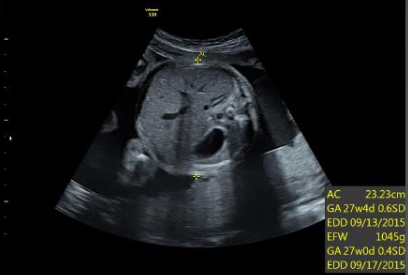
Third trimester ultrasound is usually performed in the context of mother’s clinical history and the results of previous ultrasound scans (such as those areas covered in second trimester morphology scan) and investigations, specifically to follow up the well-being status of baby.

To conduct review on baby’s anatomy including size and well-being:

  • Head – biparietal diameter (BPD雙頂直徑) and head circumference (HC頭圍).
  • Abdomen – abdominal circumference (AC腹圍).
  • Leg – femur length (FL股骨長度).
  • Arm – humerus length (HL肱骨長度).
  • In sum, to assess whether the baby is too small or too large, as there may be conditions of mother affecting the growth of the baby.

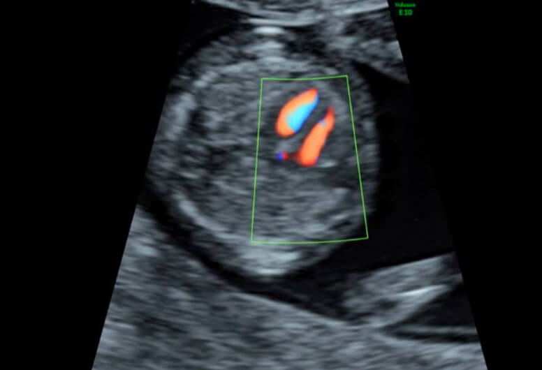
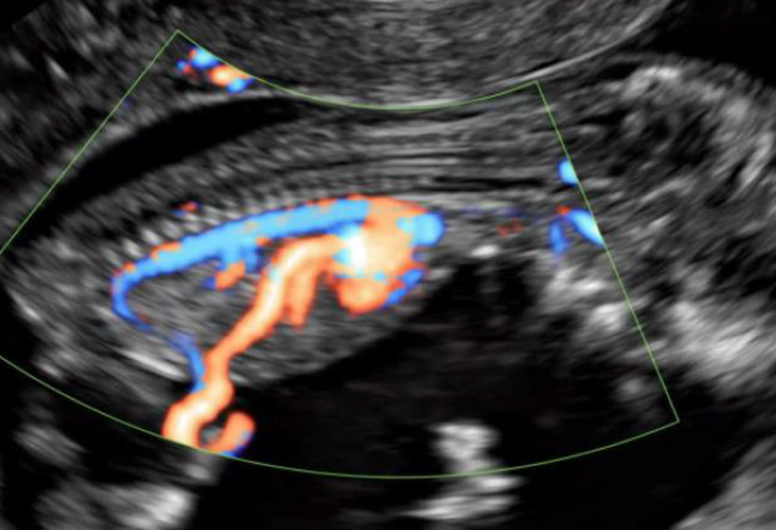
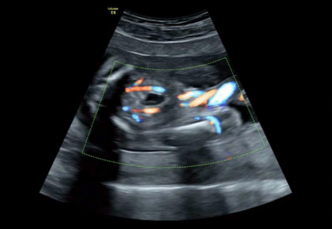
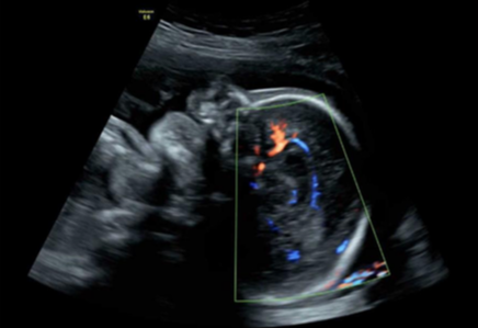
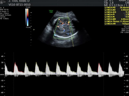
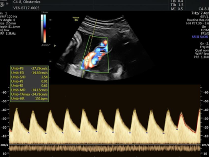
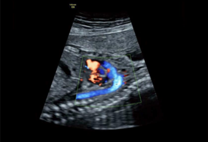
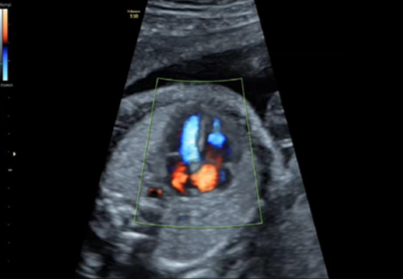
To check the baby’s heart rate and rhythm.

To review the position of the placenta, see whether there are concerns of a low-lying placenta (低位胎盤) or placenta praevia (前置胎盤) requiring intervention to prevent complications.

To assess the position of the baby, as this becomes more important when approaching delivery.

WHAT TO BE CHECKED IN THIS STAGE:

  • The length of the cervix (宮頸長度) may need checking, especially if mother has premature labour (早產), vaginal bleeding or pain. Sometimes a transvaginal ultrasound scan is needed to get a better view of the cervix. But for full term baby (足月嬰兒), the length of the cervix is not as important.
  • To conduct precise measurement on volume of amniotic fluid (羊水), which is usually expressed as the “amniotic fluid index” (AFI 羊水指數). The volume of fluid around the baby may be increased above normal (polyhydramnios 羊水過多), or is below the normal range (oligohydramnios羊水過少). Changes in the fluid volume are not always significant, but there may be possible problems with mother or baby, for example, polyhydramnios can be related to gestational diabetes (妊娠糖尿病), and oligohydramnios can be associated with small baby. The measurement of amniotic fluid volume can be conducted more frequently as required.
  • To measure blood flow in the umbilical cord (臍帶). Doppler ultrasound scan (多普勒超聲波掃描) is used to check blood flow in the umbilical artery (檢查臍動脈的血流量), sometimes blood flow in the baby’s brain and liver are also checked. They help assess the function of the placenta and the health status of baby and the Doppler ultrasound scan can be performed more frequently if required. The measurements are expressed in specific terms such as Pulsatility Index (PI), Resistance Index (RI), and Systolic/Diastolic Ratio (S/D ratio). Growth restricted baby (生長受限的嬰兒) may show progressive changes in the blood flow of these vessels. Monitoring such changes can help the doctor decides whether the baby needs to be delivered earlier.
  • Group B streptococcus screening (B組鏈球菌篩查) for mother: Vaginal and rectal swabs are taken at 35 to 37 weeks of gestation to diagnose Group B strep bacteria. Although Group B strep can be present in up to 30% of healthy mothers, it’s the leading cause of life-threatening infections in newborns and can also cause mental retardation, impaired vision, and hearing problem. Mothers who are tested positive can be treated with antibiotics during delivery to protect the baby from infection at birth.